


About the blog author: Regina Bedgood (she/her) wrote this blog post as part of Dr. Stacy Krueger-Hadfield’s Science Communication Course at the University of Alabama at Birmingham. She earned her BS in Biology with a minor in Chemistry at UAB. She is currently a PhD student in Dr. Karolina Mukhtar’s lab at UAB where she studies molecular plant biology. Specifically, she researches biotic and abiotic stress of plants using Arabidopsis thaliana as a model organism. In her spare time, Regina enjoys keeping planted aquaria, rock climbing, skateboarding, and photography. You can find her @reggieb126 on Instagram.
As a kid, did you have glow-in-the-dark stars on your ceiling? If you did, then you are familiar with something called fluorescence!
Fluorescence happens when an object or molecule absorbs light at one wavelength, then emits the light at a different wavelength. When we see glow-in-the-dark stars glow, we are observing fluorescence. Therefore, a more accurate name for glow-in-the-dark stars would be fluorescent stars, but that doesn’t sound quite as imaginative.
In biology, we often use fluorescent proteins to uncover mysteries happening inside cells. Fluorescent proteins act similarly to glow-in-the-dark stars, only much smaller. We can picture them as microscopic glow-in-the-dark stars inside cells. But how is this useful for a biologist?
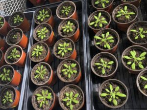
I study a small model organism called Arabidopsis thaliana. It’s a cute plant which is very important in the world of plant science (Figure 1). Specifically, I study how it responds to stress, such as a pathogen infection.
Unlike humans who have specialized immune cells, every cell can act as an immune cell in plants. When a plant cell senses a pathogen, it will quickly begin responding. The first step in the response is the movement of ions to different subcellular compartments (Figure 2).
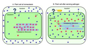
This movement of ions initiates a series of biochemical changes putting the cell in defense mode. This is happening at the subcellular level. How is it possible to observe changes happening in such a tiny space?
You guessed it, fluorescent proteins!
Fluorescent proteins can be ion sensitive, meaning they fluoresce in a different color depending on the concentration of a particular ion. They can also be molecularly tethered to specific locations within a cell, much like taping glow-in-the-dark stars to the ceiling or wall (Kesten et. al., 2019; Gjetting et. al.,2012; Martinière et. al., 2018). This means a leaf from an Arabidopsis plant can be stressed and monitored under a high-powered microscope to observe where ions are moving inside the cells.
Such fluorescent protein lines of Arabidopsis are allowing scientists to uncover aspects of plant immunity not formerly observable. Most mechanisms previously used to test pH or other ion levels in plants were invasive, meaning they damaged tissue, and were not localized to a particular area within the plant cells. Therefore, having a non-invasive mechanism for observing pH and calcium levels of subcellular compartments in real time within living tissues is incredibly exciting.
2021-05-21T11:45:52
Camera Name:
Numerical Aperture: 1.3
Refractive Index: 1.515
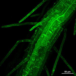
This amazing science can be understood with the same little glowing stars many of us counted on our ceilings while falling asleep as children. I will leave you with an image of the root of a pH sensitive green fluorescent protein line of an Arabidopsis seedling so you too can enjoy the mesmerizing world of fluorescent proteins (Figure 3).
References: